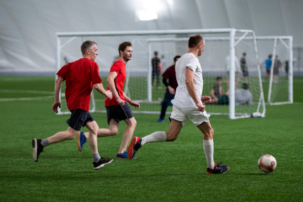
Soft tissue and tendon injuries commonly occur following sporting injuries, sudden twisting movements, falls, workplace accidents, running or jumping activities, or other forms of trauma. These injuries can affect the muscles, tendons, ligaments, and supporting structures of the hip, thigh, knee, lower leg, and ankle, often causing significant pain, swelling, weakness, instability, and difficulty walking or performing physical activity.
Some injuries occur following a sudden high-force movement or direct trauma, while others may develop due to repetitive strain, overuse, or tendon degeneration over time. Tendon ruptures can sometimes occur during explosive movements such as jumping, sprinting, or pushing off suddenly.
The severity of soft tissue and tendon injuries can vary from mild sprains or strains managed with rehabilitation and activity modification through to more significant tendon tears or ruptures that may require surgical treatment to restore stability, strength, and function.
Dr Scott Tulloch consults for a range of soft tissue and tendon injuries, which are outlined in further detail below. Treatment recommendations are tailored to the specific injury pattern, severity of the tendon or soft tissue damage, functional requirements, and overall health of each patient.
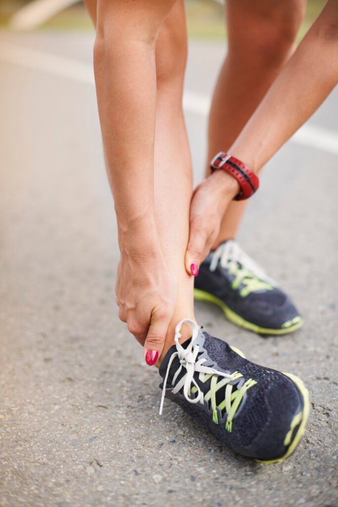
An ankle syndesmosis injury, commonly referred to as a high ankle sprain, involves injury to the ligaments that connect the two lower leg bones, the tibia and fibula, just above the ankle joint. These ligaments help maintain stability of the ankle during walking, running, and twisting movements. High ankle sprains commonly occur during sporting activities, particularly those involving sudden twisting, pivoting, or impact to the ankle, such as football, basketball, skiing, or rugby. These injuries can also occur following falls or other traumatic ankle injuries.
Symptoms often include:
Because syndesmosis injuries can sometimes occur alongside ankle fractures or other ligament injuries, prompt assessment is important. Diagnosis is typically made through a combination of clinical examination and imaging, including X-rays and, in some cases, MRI scans to assess the extent of ligament damage and ankle stability.
Treatment depends on the severity of the injury and degree of instability.
Mild injuries may be managed with:
More significant or unstable injuries may require surgical stabilisation to restore alignment and stability between the tibia and fibula.
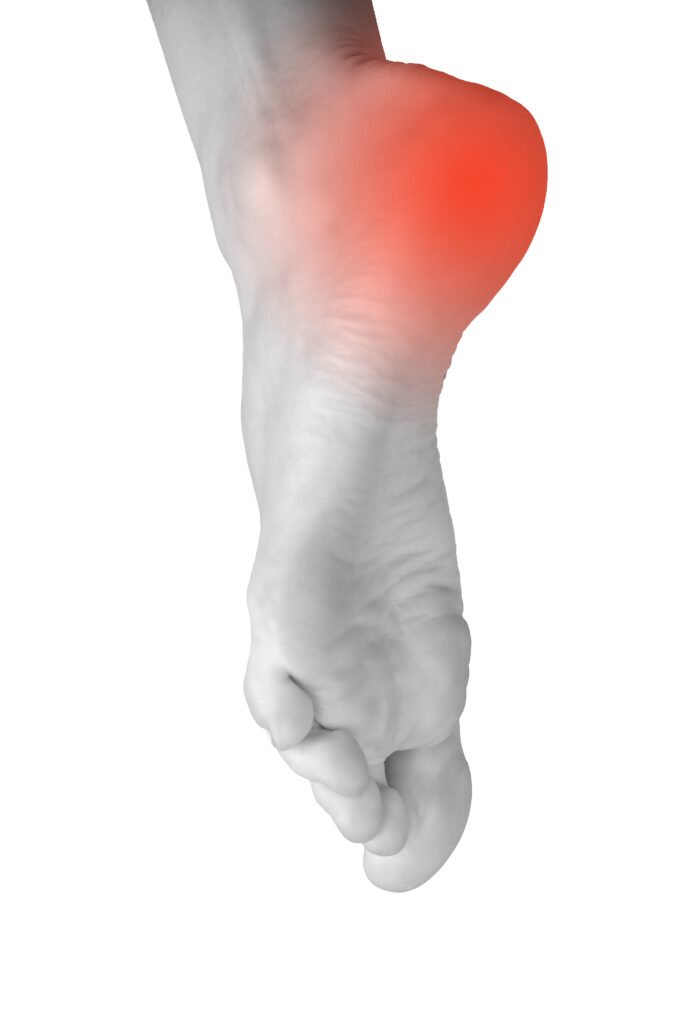
Sinus tarsi syndrome is a condition involving pain and inflammation in a small space on the outside of the ankle known as the sinus tarsi, located between the talus and calcaneus bones. This area contains ligaments, soft tissue, and nerve endings that contribute to ankle stability and movement.
Sinus tarsi syndrome commonly develops following an ankle sprain or repetitive ankle instability, although it can also occur due to overuse, altered foot mechanics, flat feet, or inflammatory conditions. Symptoms may develop gradually or persist after a previous ankle injury.
Common symptoms may include:
Diagnosis is typically made through a combination of clinical examination and imaging, which may include X-rays, ultrasound, or MRI scans to assess the surrounding ligaments, soft tissues, and joint structures.
Treatment depends on the underlying cause and severity of symptoms. Many cases can be managed with:
In some cases, injections or surgical treatment may be considered if symptoms persist despite non-surgical management.
An Achilles tendon rupture occurs when the Achilles tendon, which connects the calf muscles to the heel bone, partially or completely tears. This tendon plays an important role in walking, running, jumping, and pushing off the foot.
Achilles tendon ruptures commonly occur during sport or physical activity, particularly during sudden acceleration, jumping, pivoting, or forceful pushing movements. Some people describe feeling or hearing a sudden “pop” in the back of the ankle at the time of injury.
Common symptoms may include:
Many patients find it difficult or impossible to stand on tiptoes following the injury.
Diagnosis is typically made through a combination of clinical examination and imaging, which may include ultrasound or MRI scans to assess the extent of the tendon injury and degree of tendon separation.
Treatment depends on factors such as the severity of the rupture, activity level, tendon quality, and functional goals of the patient. Some Achilles tendon ruptures can be managed with:
In other cases, particularly in more active individuals or where there is significant tendon separation, surgical repair may be recommended to restore tendon continuity and strength.
Hamstring tendon injuries involve damage to the tendons or muscles at the back of the thigh, which help control hip extension and knee bending during walking, running, and sporting activity. These injuries can range from minor strains through to more significant partial or complete tendon tears.
Hamstring injuries commonly occur during sporting activities involving sprinting, jumping, sudden acceleration, or forceful stretching, although they can also occur following falls or other traumatic movements.
Common symptoms may include:
In some cases, patients may feel a sudden tearing sensation at the time of injury. Diagnosis is typically made through a combination of clinical examination and imaging, which may include ultrasound or MRI scans to assess the location and severity of the tendon injury.
Treatment depends on the severity of the injury, degree of tendon tearing, activity level, and functional goals of the patient. Many hamstring injuries can be managed with:
More significant tendon ruptures, particularly where the tendon has pulled away from the bone, may require surgical repair to restore strength and function.

A quadriceps tendon rupture occurs when the tendon connecting the quadriceps muscles at the front of the thigh to the kneecap (patella) partially or completely tears. This tendon plays an important role in straightening the knee, walking, climbing stairs, and standing from a seated position.
Quadriceps tendon ruptures commonly occur following a sudden forceful movement, such as jumping, landing awkwardly, stumbling, or attempting to prevent a fall. These injuries are more common in middle-aged and older adults, particularly where there is underlying tendon degeneration or medical conditions affecting tendon strength.
Common symptoms may include:
In complete ruptures, patients are often unable to perform a straight leg raise or actively extend the knee.
Diagnosis is typically made through a combination of clinical examination and imaging, which may include X-rays, ultrasound, or MRI scans to assess the extent of tendon damage and identify any associated injuries.
Treatment depends on the severity of the rupture, degree of tendon separation, activity level, and functional requirements of the patient. Partial tears may sometimes be managed with:
Complete ruptures commonly require surgical repair to restore tendon continuity and knee extension strength.
A patellar tendon rupture occurs when the tendon connecting the kneecap (patella) to the shin bone (tibia) partially or completely tears. The patellar tendon works together with the quadriceps muscles and quadriceps tendon to help straighten the knee and support walking, running, jumping, and climbing stairs.
Patellar tendon ruptures commonly occur during sporting activities or sudden forceful movements, particularly jumping, landing, or rapid changes in direction. These injuries may also occur following a fall or direct trauma to the knee. In some cases, underlying tendon degeneration or previous tendon problems can increase the risk of rupture.
Common symptoms may include:
In complete ruptures, patients are often unable to actively extend the knee or perform a straight leg raise.
Diagnosis is typically made through a combination of clinical examination and imaging, which may include X-rays, ultrasound, or MRI scans to assess the extent of tendon damage and identify any associated injuries.
Treatment depends on the severity of the rupture, degree of tendon separation, activity level, and functional requirements of the patient. Partial tears may sometimes be managed with:
Complete ruptures commonly require surgical repair to restore tendon continuity and knee function.
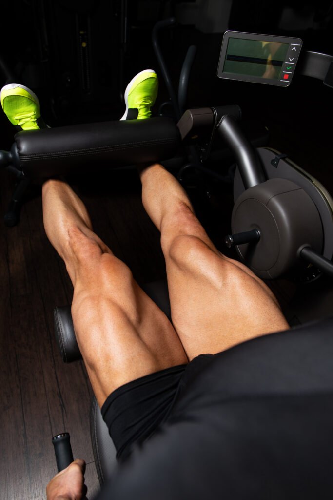
Rehabilitation is an important part of recovery and focuses on restoring movement, strength, balance, and knee stability before gradually returning to higher-demand activity.