Osteochondritis Dissecans (OCD)
A condition affecting the bone and cartilage of the knee, leading to pain, swelling, and joint symptoms
Osteochondritis dissecans (OCD) is a condition that affects the bone and overlying cartilage within a joint, most commonly the knee. It occurs when a small area of bone beneath the cartilage loses its normal blood supply, which can affect the stability of both the bone and the cartilage surface.
Over time, this area may become weakened, and in some cases, a fragment of bone and cartilage can begin to separate from the surrounding tissue. This can lead to symptoms such as pain, swelling, stiffness, and changes in joint movement, including catching or locking sensations.
Early assessment is important to determine the stage and stability of the condition, as this helps guide appropriate management and supports long-term joint health and function.
What is osteochondritis dissecans (OCD)?
Osteochondritis dissecans (OCD) is a condition that affects the bone and overlying cartilage within a joint, most commonly the knee. It occurs when a small area of bone beneath the cartilage develops reduced blood supply, which can weaken the bone and affect the stability of the cartilage surface above it. As the condition progresses, this area may become less stable, and in some cases a fragment of bone and cartilage can begin to separate from the surrounding tissue. This may remain in place or, in more advanced cases, become partially or fully detached, which can interfere with normal joint movement.
OCD can vary in severity. In earlier stages, the bone and cartilage may remain intact but weakened. In later stages, there may be loosening or displacement of a fragment, which can contribute to symptoms such as pain, swelling, and mechanical symptoms within the joint.
The condition is more commonly seen in children and adolescents whose bones are still developing, although it can also occur in adults. Early diagnosis is important, as the stability of the affected area helps guide treatment decisions and potential for healing.
Common causes of osteochondritis dissecans
The exact cause of osteochondritis dissecans (OCD) is not always clearly defined, but it is thought to develop due to a combination of repetitive stress and changes in blood supply to the bone beneath the cartilage. In many cases, OCD is associated with repetitive loading of the joint, particularly during activities that involve running, jumping, or high levels of impact. Over time, this repeated stress may affect the underlying bone, leading to changes in its strength and stability.
Common contributing factors include:
- Repetitive stress or overuse, particularly in active children and adolescents involved in sport
- Reduced blood supply to a small area of bone, which can affect its ability to remain healthy and support the cartilage above
- Microtrauma, where repeated small injuries accumulate over time rather than a single significant event
- High levels of physical activity during growth, when the bone is still developing
In some cases, OCD may also be influenced by:
- Joint alignment or biomechanics, which can affect how forces are distributed through the knee
- Genetic or individual factors, which may affect bone development and resilience
Unlike many other joint injuries, OCD often develops gradually, without a single identifiable injury. Understanding the contributing factors can help guide both treatment and activity modification, particularly in younger, active individuals.
Symptoms of osteochondritis dissecans
Symptoms of osteochondritis dissecans (OCD) can develop gradually and may vary depending on the stage and stability of the affected area. In the early stages, symptoms may be mild and related to activity, but they can become more noticeable as the condition progresses.
Common symptoms include:
- Knee pain, often worse with activity such as running, jumping, or sports
- Swelling, which may come and go depending on activity levels
- Stiffness, particularly after periods of rest
- Reduced range of motion, making it difficult to fully bend or straighten the knee
As the condition advances, particularly if a fragment becomes unstable, you may also experience:
- Catching or locking sensations within the joint
- A feeling of the knee not moving smoothly
- Intermittent sharp pain, especially with certain movements
Symptoms can fluctuate, often worsening with activity and improving with rest. In some cases, symptoms may persist even with reduced activity if the affected area becomes unstable.
Risk factors for osteochondritis dissecans
A number of factors may increase the likelihood of developing osteochondritis dissecans (OCD), particularly in individuals who are physically active or still growing.
Common risk factors include:
- Age and skeletal maturity, with OCD more commonly seen in children and adolescents whose bones are still developing
- High levels of sporting activity, especially activities that involve running, jumping, or repetitive impact
- Repetitive stress on the joint, which may affect the underlying bone over time
- Rapid growth phases, where changes in bone development may influence how the joint responds to load
Additional factors that may contribute include:
- Joint alignment or biomechanics, which can affect how forces are distributed through the knee
- Previous minor joint injuries, which may contribute to repeated stress in a specific area
- Genetic or individual factors, which may influence bone health and development
In many cases, OCD develops due to a combination of these factors rather than a single cause.

Treatment options for osteochondritis dissecans
Management of osteochondritis dissecans (OCD) is guided by the stage of the condition, stability of the affected area, your age, and activity level. Treatment may be non-surgical or surgical, depending on whether the bone and cartilage remain stable. Non-surgical treatment options are often considered in younger patients or when the affected area is stable.
These may include:
- Activity modification, reducing high-impact activities such as running and jumping
- A period of rest or protected weight-bearing, in some cases
- Physiotherapy, to maintain strength and joint movement while protecting the area
- Regular monitoring, including follow-up imaging, to assess healing
In many cases, particularly in growing individuals, stable lesions may improve over time with appropriate management. If the affected area is unstable, or if symptoms persist despite non-surgical treatment, surgical options may be considered.
These can include:
- Fixation of the fragment, where the bone and cartilage are secured back in place
- Removal of loose fragments, if the tissue is no longer viable
- Cartilage repair or restoration techniques, aimed at improving the joint surface
The most appropriate treatment depends on factors such as the size and location of the lesion, joint stability, and your functional needs.
When to seek medical advice for osteochondritis dissecans
It is appropriate to seek medical advice if you are experiencing persistent knee pain, swelling, or changes in joint movement, particularly if symptoms are ongoing or affecting your ability to stay active.
You may benefit from an assessment if you notice:
- Ongoing knee pain, especially during or after activity such as running or sport
- Swelling that recurs or does not settle with rest
- Stiffness or reduced range of motion
- A feeling of catching, clicking, or locking within the knee
- Symptoms that are progressively worsening or limiting activity
In children and adolescents, early symptoms may be mild and easily overlooked. If a young person involved in sport develops persistent knee pain, it is important to have this assessed, particularly during periods of rapid growth. It is especially important to seek review if there are mechanical symptoms, such as locking or the knee feeling stuck, as this may indicate that the affected area has become unstable. Early assessment can help determine the stage of the condition and guide appropriate management, which may improve the likelihood of successful treatment and support long-term joint health.
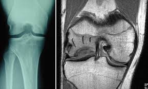
Dr Scott Tulloch will perform a comprehensive assessment, which may include imaging such as X-rays or MRI, to evaluate the condition and discuss the most appropriate treatment options based on your individual needs.